
Background
Amyotrophic Lateral Sclerosis (ALS) is a fatal disorder, which results in the progressive loss of motor neurons. Motor neurons are the cells that communicate information from the movement centre of the brain, through the spinal cord to our muscles. Patients with ALS show great variability in the age they are diagnosed, the types of symptoms they experience, and how long they will live with the condition. This makes it difficult to give patients an accurate prediction of quality of life and life expectancy. Most of the research into ALS has focused on the motor neurons, however there is growing evidence to indicate other cells within the central nervous system (CNS) also contribute to the disease. These include glial cells, which support the functioning of neurons, and vascular cells which make up our blood vessels.
This study aimed to determine which cells of the central nervous system contribute to disease, and to look at how gene expression changes over the course of disease. Gene expression is the process by which information from specific parts of our DNA, called genes, is used to make proteins. Cells require proteins to perform all their functions.
Why is the study important?
Having a better understanding of the contributions of other cells within the CNS and the changes in cell behaviour that take place early in disease will help to provide patients with more accurate prognosis information. Improving understanding of early events in disease could provide new targets for the development of treatments.
What did the authors do and what were the results?
First, gene expression data gathered from post-mortem human spinal cord was analysed to determine which types of cells change their behaviour in ALS, and therefore play a role in disease. To do this a data analysis method called expression-weighted cell-type enrichment (EWCE) was employed. This method compares the gene expression data from individual cells of people with ALS to the average cell expression data from healthy cells, determining the important changes in cell behaviour.
This revealed an increase in the activity of glial cells and a decrease in activity of neurons. This could be the result of motor neuron death. Interestingly, vascular cells showed an increase in their activity, particularly a group of cells called perivascular fibroblasts. These cells help to support the blood vessel, keeping them strong and resistant to damage.
As post-mortem tissue can only give information about disease at the time of death, mouse models of ALS were also used. These mice have genetic mutations, which cause ALS in humans. These mice develop a condition like ALS. These mice were used to examine how gene expression changes before symptoms develop and how gene expression changes over the course of disease.
In the mouse models of ALS, the changes in gene expression were very similar to that seen in humans. Increased activity of vascular and glial cells as well as decreased neuron expression. Perivascular fibroblasts showed the biggest change in their gene expression of any cell type. These were the first type of cell to change their activity, before the mice started to show symptoms. This suggests these types of cells play an important role in the progression of ALS, and potentially the onset of symptoms. Perivascular fibroblasts are a relatively newly discovered cell, and their role in ALS has not yet been examined.
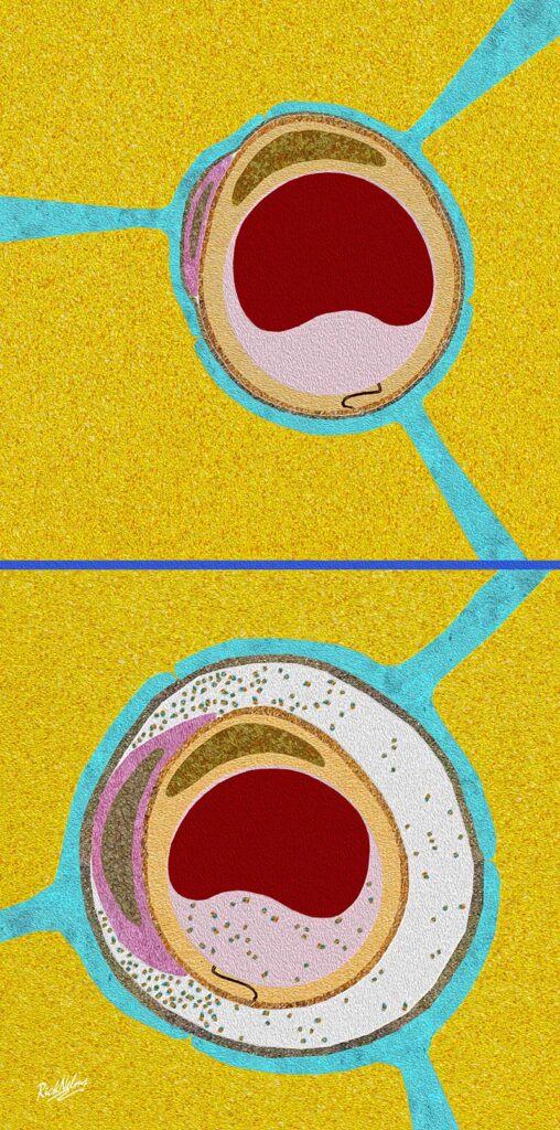
Next the researchers used a technique called immunohistochemistry (IHC) to determine the presence of proteins in sections of tissue. This method involves using proteins called antibodies that attach to the protein of interest. Dyes can then be attached to the antibodies allowing visualisation of the protein of interest in the tissue. The researchers stained sections of spinal cord from people with ALS and from people without any neurological disease (known from here as healthy controls) for two proteins known to be produced by perivascular fibroblasts – SPP1 and COL6A1. In people with ALS, the perivascular fibroblasts produced more SSP1 and COL6A1 than in the healthy controls. These proteins were mostly found in perivascular spaces. These spaces are the small gaps between the blood vessel and the surrounding brain tissue. In the spinal cord tissue of people who had ALS these spaces were much larger compared to the spinal cord from those who hadn’t had ALS. In the ALS mouse models, these enlarged perivascular spaces were observed before the mice started to show symptoms. This suggests that the structure of blood vessels changes very early in disease.
The brain and spinal cord sit the fluid called cerebrospinal fluid. Perivascular spaces help to drain cerebrospinal back into blood vessels. Researchers wanted to see whether SSP1 and COL6A1 would be detectable in patient blood. If so, this could be used as a method of tracking the progression of disease in patients.
Blood collected from 574 ALS patients was analysed to determine the amount of SSP1 and COL6A1 present. Higher levels of SSP1 and COL6A1, at the time of diagnosis, were associated with a shorter survival time. Having continuously increasing amounts of SSP1 in blood, was a better predictor of shorter patient survival, than already established predictors of patient survival. These predictors included a particularly aggressive form of ALS known as bulbar onset ALS, in which symptoms first begin in the face and neck, and levels of neurofilament protein in cerebrospinal fluid which is currently one of the best predictors of survival .
What do the finding mean going forward for people with the disease?
This study has shown that changes in perivascular fibroblast cells and blood vessel structure are important events in early ALS. By developing a better understanding of what causes these changes and what effect they have on the brain, researchers can develop a better understanding of what potentially causes ALS and provides new areas for treatment development. Levels of the SSP1 protein present in blood, could help to give patients more information about their prognosis following diagnosis.