
Background
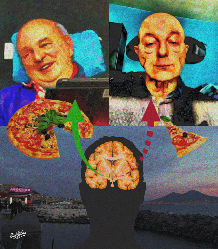
Amyotrophic lateral sclerosis (ALS) is the most common form of motor neurone disease (MND). In ALS, motor neurones develop damage over time and stop functioning properly, causing muscle weakness and eventually paralysis. Previous studies on people with ALS and animal models show that weight loss occurs as ALS progresses, with faster weight loss linked to faster ALS progression. This suggests that people with ALS are hypermetabolic, or they burn energy at a faster rate. Increasing calorie intake may help in slowing ALS progression. Recent studies have also shown that appetite changes in people with ALS, which may also impact weight loss. This study aimed to develop a better understanding of how ALS impacts appetite control in the brain.
Why is the study important?
We may be able to slow ALS progression by increasing food intake in people with ALS. If the brain’s ability to control appetite is disrupted in ALS, this may be more difficult to achieve. This study looks in more detail at appetite-controlling brain regions in ALS, to see if anything is disrupted and could be a therapeutic target.
What did the authors do and how did they do it?
The authors studied brain samples from 17 people with ALS and compared them to samples from 13 people without ALS. They obtained brain samples from autopsies from previous studies. They looked at markers of damage usually seen in motor neurones in ALS, in a region of the brain that controls appetite, called the lateral hypothalamic area (LHA).
To try and investigate the impact of these changes , the authors studied mice with genetic changes that cause ALS. They studied the same brain region to see if the same neurones were damaged in the ALS-mice. They also used an intracerebroventricular shunt, a device that allows injections into the fluid surrounding the brain to ensure drugs are delivered to the brain effectively. They used this device to inject the appetite controlling hormone MCH, which is produced by the damaged neurones identified earlier, to try and replace any lost hormone. The authors monitored the body weight and food intake of the ALS- mice that were receiving these hormone injections around their brains. The authors also looked at respiratory exchange ratio (RER), a measurement that tells us whether an animal is mostly using fat or carbohydrates as a fuel source from the diet to produce energy. This was done to see if injecting the hormone caused the mice to change the fuel they were using from their food.
What are the results?
ALS- mice had fewer neurones that produce the appetite hormone MCH, suggesting their brains have less ability to control appetite. When mice were injected with MCH, they stopped losing weight, ate more food and started to use more carbohydrates instead of fat for energy.
In brain tissue from 17 people with ALS, the markers of neurone damage called lipofuscin and pTDP-43 were studied. These markers are usually increased in motor neurones in people with ALS. The authors found these markers are also increased in MCH-producing neurones, showing they may become damaged in ALS in a similar way to motor neurones. There were fewer appetite-increasing neurons in brain samples from people with ALS. Many of these remaining neurons also appeared to be dying.
What do the finding mean going forward for people with the disease?
This study tried to gather more information on appetite changes in models of ALS, to see if they have an effect on weight loss seen during the disease. Targeting this weight loss with injections of the hormone MCH may help increase appetite, reduce weight loss and slow ALS progression.
In the mouse models of ALS, the authors did not look at whether injection of the appetite-increasing hormone around the brain could extend survival or slow the worsening of symptoms in the mice. This would be a useful follow up study to confirm that these hormone injections are effective in helping people with ALS.