
7,8-Dihydroxyflavone protects neurons against oxygen-glucose deprivation induced apoptosis and activates the TrkB/Akt pathway
Summary by Shivani
Background:
A stroke occurs when the blood supply to the brain is either reduced or stopped. The most common type of stroke is an ischemic stroke, caused due to a blood clot in the blood vessels that supply blood to the brain (shown in figure 1).
Like all organs in the body, the brain relies on nutrients (such as glucose, or sugar) and oxygen, delivered via blood, for it to carry out its functions. An adequate supply of glucose and oxygen is essential to maintain the health of neurons, the cells that send and receive signals in the brain. Therefore, when a stroke occurs due to inadequate blood supply, neurons in the brain are deprived of oxygen and glucose. This process, called oxygen and glucose deprivation (OGD) causes neuronal injury and death post-stroke.
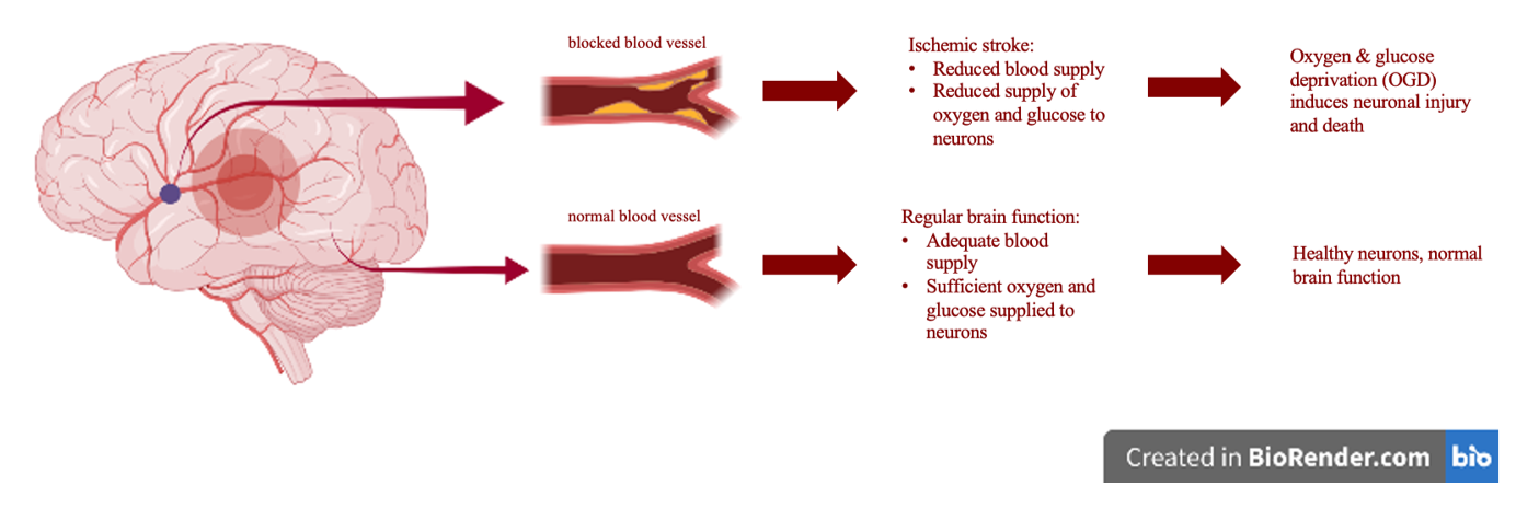
Figure 1. Mechanism of Ischemic Stroke.
All cells, including neurons, contain microscopic substances called molecules. These enable the cell to carry out various functions by interacting with one another. One such molecule, called brain-derived neurotrophic factor (BDNF), plays a key role in the survival of neurons. It exerts its effects by activating another molecule, called TrkB, which in turn activates another molecule, which activates another. Such a series of events is called a molecular cascade or a signalling pathway. The signalling pathway that is activated by BDNF is called the BDNF/TrkB/Akt pathway, or simply the TrkB/Akt pathway.
The TrkB/Akt pathway has been linked to neuronal survival and is thought to protect the neurons against oxygen and glucose deprivation (OGD). Since OGD is thought to be the mechanism of neuronal death after stroke, activating this pathway might provide a method to improve the health of neurons and facilitate their recovery post-stroke.
Research has shown that the TrkB/Akt pathway can be activated artificially by an experimental drug called 7,8-dihydroxyflavone (7,8-DHF), which mimics the effects of BDNF. Some studies have shown the beneficial effects of 7,8-DHF in other neurodegenerative disorders, but its potential benefit has not been explored in the context of ischemic stroke.
Why is this study important?
Ischemic strokes account for about 85% of strokes worldwide. They are a leading cause of long-term disability and death worldwide. Therefore, developing new treatment strategies for ischemic stroke is extremely important. This study provides insight into the mechanisms that are affected at the cellular level during ischemic stroke. These pathways offer potential targets for the development of new therapies that may eventually benefit the patient community.
What did the authors do and how did they do it?
Using animal cells to model diseases in humans is a common practice to test the effects of experimental drugs, whose effects are unknown, as these cannot be tested directly in humans. The use of animals in medical research is a highly regulated process with many ethical, legal, and moral considerations.
In this study, the researchers used neurons derived from rats to investigate whether 7,8-DHF can protect neurons from OGD-induced injury.
The researchers grew the rat neurons in conditions lacking oxygen and glucose, so as to mimic the OGD that accompanies stroke in people. A subset of these cells was treated with 7,8-DHF to determine whether it had potential beneficial effects as compared to the control cells, that were untreated. A range of tests were performed to confirm that OGD caused the death of neurons, and to subsequently determine whether 7,8-DHF treatment could aid neuronal recovery and survival via the activation of the TrkB/Akt pathway.
What are the results?
To achieve the aims set out in this study, the researchers used a range of tests to determine the effects of OGD and 7,8-DHF treatment on neurons. These tests are aimed at detecting markers for cell death, cell survival and activation of the TrkB/Akt pathway. The tests were carried out in a test group of neurons – that were treated with 7,8-DHF after OGD, and a control group of neurons – that were subjected to OGD and left untreated. Comparing the results from the test and control group enabled the researchers to confirm that the effects observed after performing the experiments are indeed due to the treatment of 7,8-DHF and not any other external factor.
The researchers found that OGD induced the death of neurons. They also found that the cells treated with 7,8-DHF showed fewer markers of cell death, thus suggesting that 7,8-DHF treatment protected the neurons from OGD-induced cell death.
The researchers also found that the TrkB/Akt pathway was less active in the untreated neurons, and that this reduced activity was reversed in the neurons treated with 7,8-DHF. This is in line with other studies that have investigated 7,8-DHF as a potential therapy in other neurodegenerative disorders.
Taken together, these results suggest that 7,8-DHF treatment can improve cell survival by activating the TrkB/Akt pathway post-stroke.
What do these findings mean going forward for people with the disease?
These findings provide insights into the links between the BDNF/TrkB/Akt signalling pathway and neuronal survival in the context of stroke. The findings of this study suggest that by influencing the activity of the BDNF/TrkB/Akt pathway, we might alleviate the damage caused to cells by OGD after ischemic stroke. This might allow for the development of new effective treatment options for patients post-stroke.
This study can be found at: https://doi.org/10.7717/peerj.12886.
Original paper title: 7,8-Dihydroxyflavone protects neurons against oxygen-glucose deprivation induced apoptosis and activates the TrkB/Akt pathway
Author list: Qinxiang Zhou, Hao Tang, Dingqun Bai and Yuhan Kong
Publication details including date of publication: This paper was published on 15th February 2022 in the journal PeerJ.
Citation:
Zhou Q, Tang H, Bai D, Kong Y. 2022. 7,8-Dihydroxyflavone protects neurons against oxygen-glucose depriva- tion induced apoptosis and activates the TrkB/Akt pathway. PeerJ 10:e12886 http://doi.org/10.7717/peerj.12886